Determining the cell number by measuring transmittance

The increasing number of yeast cells (Saccharomyces cerevisiae), increases the turbidity of the sample. Increasing turbidity decreases the intensity of light passing through the sample. This affects the transmittance measured in the given sample. Increasing cell number decreases measured transmittance.

In order to determinate the number of yeast cells using transmittance, the sample must be stabilized and the calibrating curve must be made.

The yeast cells in the sample are reproducing; their numbers grow in time. The cell number and the transmittance should be determinate in the same sample. The sample 's characteristics must not be changed during the process of analysis. If the sample is heated at 80°C for 3 minutes, the yeast cells are inactivated but not disintegrated. Their number doesn’t change and the sample can still be analyzed.

The calibrating curve shows how the cell number affects measured transmittance. The number of cells in the 1 mL of the sample is enormous (20.24·109 cells/mL). In the calibrating curve the number of cells should be given as ln cell number (natural logarithm of the number of yeast cells in 1 mL of sample).

The cell number in 1 mL of the sample can be determined with a hemocitometer and a light microscope.

Measuring transmittance and counting cells to create the calibration curve


How to prepare the initial sample

  1. using fresh bakery yeast

    Mix 1 g of fresh bakery yeast and 100 mL of sterilized water solution (0.9 % NaCl). Stir until the liquid is homogenized. Whilst stirring, heat the given liquid with cells to 80°C and keep it at this temperature for 3 minutes. This is the initial sample.
  2. Using dry (exsiccated) bakery yeast

    Heat 200 mL of sterilized distilled water to 30°C and dissolve 2 g of glucose (C6H12O6) in it. Mix 14 g of dry (exsiccated) bakery yeast with the prepared glucose solution and wait until the yeast cells revitalize. This is when bubbles of carbon dioxide begin to rise. When the yeast cells in the liquid are revitalized, heat the liquid with cells to 80°C and keep it at this temperature for 3 minutes. This is the initial sample.

How to prepare diluted samples

Set up five labelled clean and sterilized tubes. In the first tube transfer 10 mL of well stirred initial sample with a clean and sterile pipette. All the samples must be well stirred before transferring them into test tubes. Using a pipette transfer 1 mL of well stirred initial sample into the second tube and add 9 mL sterilized culture media (0.9 % NaCl or glucose solution). Stir well. This sample is 10-1 diluted.

Using a pipette transfer 1 mL of the sample from the second into the third tube and add 9 mL sterilized culture media. Stir well. This sample is 10-2 diluted.

Repeat the action two more times until five samples have been made:

  • initial sample
  • 10-1 diluted sample
  • 10-2 diluted sample
  • 10-3 diluted sample
  • 10-4 diluted sample

Before each transfer of a sample, the tube you take the sample from must be well shaken.

The solution used for the dilution of the samples must always be taken from the liquid phase of the given sample.

Counting cells in the diluted samples

Number of cells in 1 mL of sample could be determined using a hemocitometer and a light microscope. A hemocitometer is used to count cells in a small amount of liquid sample. The number of cells in 1 mL of sample is calculated using the result. In order to get accurate results the cell number in the sample must not be too big or too small. It is impossible to count cells, if their number is too big. Such a sample should be diluted before counting.

Equation used to calculate number of cells in 1 mL of sample:

Cell number/1 mL sample = average cell number · 4 · 106 · R

Average cell number is calculated by dividing the number of cells in all hemocitometer squares with the number of squares.

R sample's dilution is used to calculate the number of cells in the initial sample. If the sample's dilution is 10-2, the number 102 is used in the equation. It is necessary because the dilution made the cell number a hundred times smaller.

We have to count cells in only one of the diluted samples, because the equation makes it possible to calculate the number of cells in the initial sample.

Each sample is ten times more diluted than the previous sample. It means that each sample contains ten times less cells than the previous sample.

If the number of cells in the initial sample is known, the number of cells in each sample can be calculated.

The number of cells in the 1 mL of the sample is enormous (20.24·109 cells/mL). In the calibrating curve the number of cells should be given as ln cell number (natural logarithm of the number of yeast cells in 1 mL of sample).

Transmittance measured in diluted samples

Fill five holes in the blister with well stirred samples. Fill each sample in one hole. Fill the sixth hole with sterilized culture media. Blister is filled as shown:

Blister 's hole

1

2

3

4

5

6

Content

initial sample

10-1 diluted sample

10-2 diluted sample

10-3 diluted sample

10-4 diluted sample

sterilized culture media

Sample volume

250 µL

250 µL

250 µL

250 µL

250 µL

250 µL

Put the filled blister on a shaker to prevent cell gathering on the bottom of the hole. Set the shaker on 320 RPM (rotations per minute). Blister should be left on the shaker, until the spectrometer is ready to use.

Get the spectrometer ready and turn on the blue LED light at the highest intensity.

Put the filled blister on spectrometer. Set the transmittance measured at the sterilized culture media on 100. 0 %. After that measure the transmittance of other samples. If measuring lasts too long, the blister should be set on the shaker again, because the cells will begin to gather on the bottom of the blister hole. This affects measuring results.

Write all the measured and calculated results in the given table.

sample

Nr. of cells/mL

ln cell nr.

T

A

initial


10-1


10-2


10-3


10-4


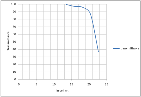
These results are used to create the graph of the calibration curve which shows how changing ln cell number affects transmittance.

Measurements used to determine the cell number
in unknown sample

How to use the calibrating curve

The given sample with the unknown number of yeast cells must be heated (80°C for 3 minutes) in order to prevent yeast cells from reproducing.

The transmittance of the stabilized sample is then measured.

If measured transmittance is lower than the lowest transmittance in calibrating curve, the sample must be diluted ten or hundred times (ten times: 1 mL sample + 9 mL dilution liquid).

If measured transmittance is higher than the highest transmittance in the given calculating curve, the calculating curve must be made again. The cell number in the initial sample of the given calculating curve is too low. The mass of bakery yeast used for the initial sample must be decreased.

The graph of the calibration curve shows how the number of cells (x) affects transmittance (y). Mark the measured transmittance (y) and read the ln of cell number (x). Calculate the number of cells/mL in the sample using the result read from the graph (ln cell nr./mL).

If the sample was diluted before this transmittance was measured, the dilution rate must be calculated in.

The liquid phase of sample can be coloured or its turbidity can be higher than water 's. It happens when apple, grape or any other fruit juice or culture media is used as the liquid phase of the sample.

The liquid that makes out the liquid phase of the sample must be used for diluting the samples. The spectrometer must also be calibrated with the same liquid.

Examples : Determining yeast (Saccharomyces cerevisiae) cell number using spectrometer measured transmittance

Example 1:

Table 1: How the number of yeast cells in samples affects transmittance measured in the same samples

sample/dilution

cell nr. /mL

ln cell nr./mL

T

A

initial

20.24 . 109

23.7

42.7

0.3696

10-1

20.24 . 108

21.4

84.6

0.0726

10-2

20.24 . 107

19.1

95.6

0.0195

10-3

20.24 . 106

16.8

95.3

0.0209

10-4

20.24 . 105

14.5

98.3

0.0075

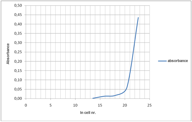
Evaluation of the results in table 1 shows that decreasing number of cells increase transmittance and decrease absorbance.

Transmittance is related to absorbance as described by the equation:

A = –log T

Graph 1: How ln cell nr./mL affects transmittance

Graph 2: How ln cell nr./mL affects absorbance

Example 2:

Table 2: How the number of yeast cells in samples affects transmittance measured in the same samples

sample/dilution

cell nr./mL

ln cell nr./mL

T

A

initial

7.23 . 109

22.7

36.7

0.4353

10-1

7.23 . 108

20.4

87.1

0.0599

10-2

7.23 . 107

18.1

96.0

0.0177

10-3

7.23 . 106

15.8

97.1

0.0128

10-4

7.23 . 105

13.5

99.6

0.0017

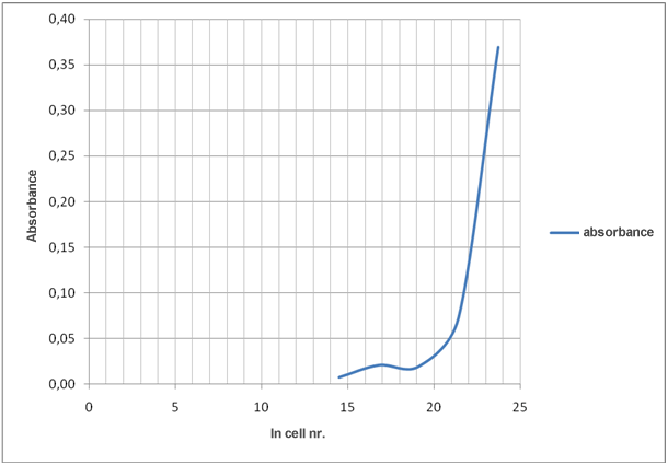
Evaluation of the results in the table 1 shows that decreasing number of cells increase transmittance and decrease absorbance.

Transmittance is related to absorbance as described by the equation:

A = –log T

Graph 3: How ln cell nr./mL affects transmittance

Graph 4: How ln cell nr./mL affects absorbance


Prepared by:
Alma Kapun Dolinar